Dental X-Ray
X-rays are an essential diagnostic procedure in dentistry. They allow dentists to detect pathological changes in the teeth, jaw, and surrounding structures early on, which are often invisible to the naked eye. The radiation exposure from modern devices is comparatively low. Nevertheless, their use should always be justified and kept as low as possible.
Intraoral X-rays
These are taken in the mouth and provide high-resolution images of individual teeth or small groups of teeth.
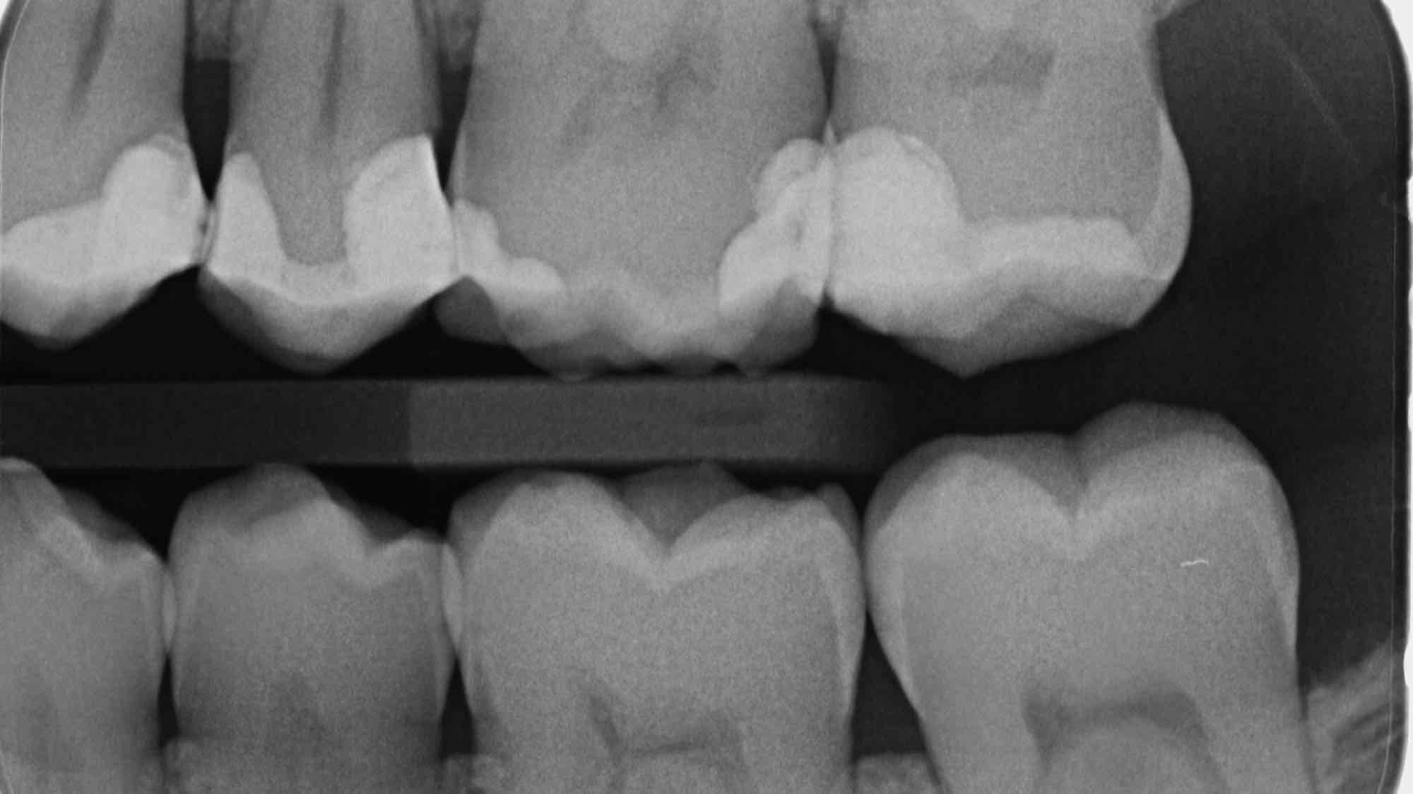
- Bitewing radiograph: Visualizes the crown areas of the upper and lower jaw simultaneously – ideal for diagnosing caries in the interdental space.
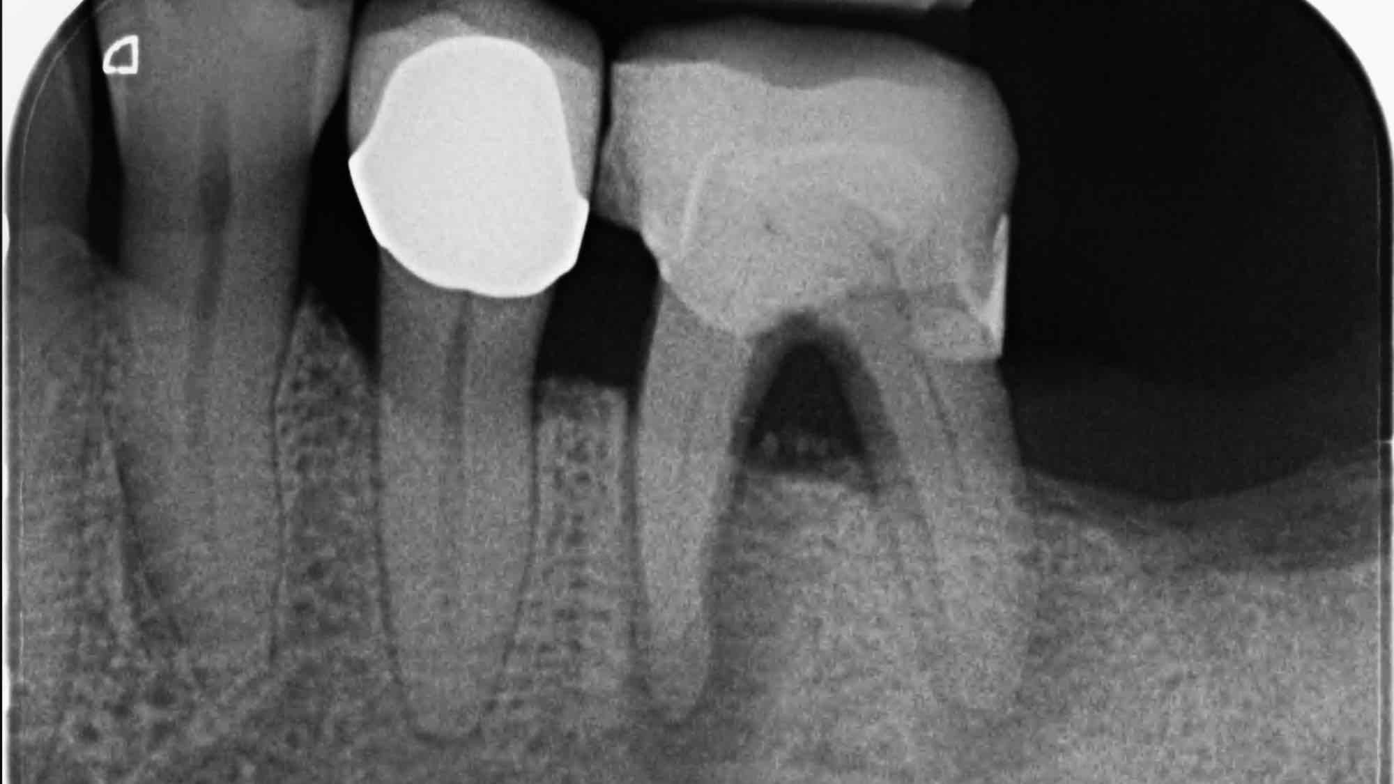
- Periapical radiograph: Shows the entire tooth, including the root tip and surrounding bone – important in cases of inflammation or root canal treatment.
Extraoral X-rays
These images are taken outside the mouth and show larger structures.
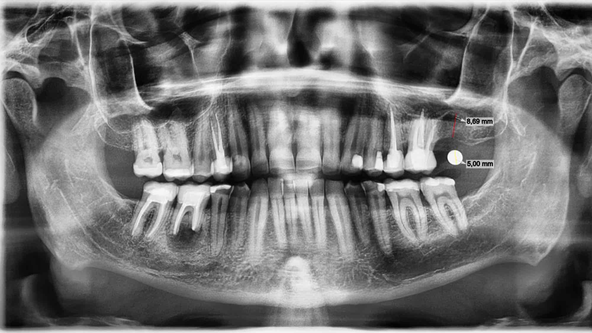
- Panoramic tomography (orthopantomography, OPG): Complete overview of all teeth, jawbones, temporomandibular joints, and adjacent structures.
- Lateral cephalogram (FSC): Lateral skull profile – primarily used in orthodontics for growth and position analysis.
- Digital cone beam computed tomography (DVT): 3D X-ray image with high detail – helpful for implant planning, complex surgical procedures, or determining the position of impacted teeth.
Indications for dental X-rays
An x-ray is usually only taken if there is a specific suspicion of a change requiring treatment or to plan a procedure. The most common indications include:
- Caries diagnosis, especially in the interdental spaces
- Checking root canal fillings or after surgical procedures
- Determining the position of displaced or impacted teeth (e.g., wisdom teeth)
- Examination of the jawbone in cases of tooth loosening or periodontitis
- Implant planning
- Fracture diagnosis after accidents
- Tumor diagnosis or exclusion
Regular x-ray checkups are also part of preventive care for certain pre-existing conditions or during orthodontic treatment.
Side effects and radiation risks
Modern X-ray machines operate with very low radiation doses, especially with digital procedures. Nevertheless, radiation exposure is considered potentially harmful to health, especially with frequent use. Potential risks:
- Cellular changes: X-rays can damage cells, which in rare cases can lead to mutations.
- Cumulative risk: Repeated X-rays over time slightly increase the overall risk.
- Sensitive groups: Children, pregnant women, and young adults are considered more sensitive to radiation and should only be X-rayed if strictly indicated.
Radiation exposure is very low thanks to modern technology, but it should still be used responsibly.
What you should do
If you want to join the winning side, then visit your dentist regularly for preventative care. They’ll also explain your weaknesses when brushing your teeth (dentists have these too!) and what you can do better. And this won’t be explained to you by a health column in the newspaper, but by someone who knows the subject inside out!
Author: drw
If you want to know more about the topic, I recommend the following links:
A very good presentation of dental x-rays from the University of Michigan:
https://www.dent.umich.edu/patient-care/dental-x-rays
Example of a panoramic tomography image
Example of a periapical dental film
Example of a bitewing radiograph


